Imaging Dentistry
-
“Professional and systematic diagnosis system that reads various dental diseases, safe treatment”
Introduction
The Department of Imaging Dentistry is one of the clinical departments of dentistry that diagnoses various diseases occurring in the teeth, oral cavity, jaw, face and neck areas through various medical images.
Intraoral radiography
Oral radiographers are mainly used for diagnosing dental caries, periodontal disease, apical disease, fractured teeth after trauma, and tooth eruption abnormalities.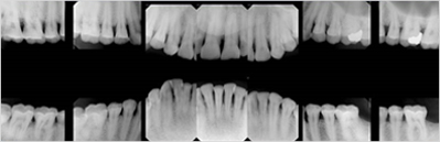
Panoramic radiography
It is mainly used for general evaluation of teeth and surrounding tissues, evaluation of tooth development, and evaluation of extensive lesions in the jawbone.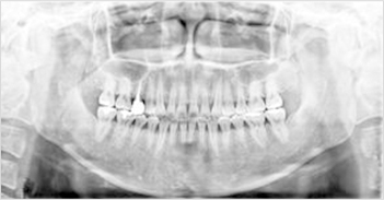
extraoral radiography
It is mainly used to evaluate lesions or traumas of the maxilla and facial bones, sinusitis odontogenic, and growth of the skull.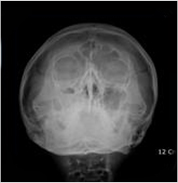
Temporomandibular joint radiograph
It is used for the diagnosis of patients with temporomandibular joint disorder and is useful for evaluating the pathological condition of the temporomandibular joint, such as bone changes in the articular surface and the positional relationship between bone structures.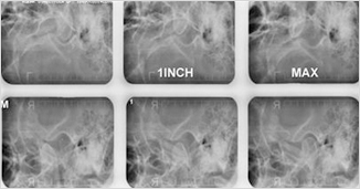
salivary gland test
Salivary gland angiography is a procedure to examine the condition of the salivary glands by injecting a drug called a radiopaque contrast agent into the salivary glands when salivary gland disease is suspected.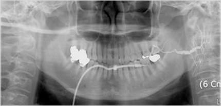
Ultrasound examination
A cross-sectional image is obtained by sending ultrasonic waves to specific organs of the human body using an ultrasonic probe (probe) and reconstructing the ultrasonic waves reflected back to the tissue with a computer. Lymph nodes, postoperative edema, hematomas, and salivary gland abnormalities can be identified.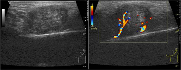
Cone Beam Computed Tomography (CBCT) for dental use only
Because dental-exclusive CT can reproduce 3D images, it is mainly used for more accurate diagnosis of dental implant procedures and temporomandibular joint disorders. In particular, the dental CT installed in the department not only takes much shorter imaging time compared to general computed tomography, but also has only 1/30 of the radiation exposure. Also, the image quality is very good, so it is very useful for precise diagnosis of various diseases occurring in the oral and maxillofacial area.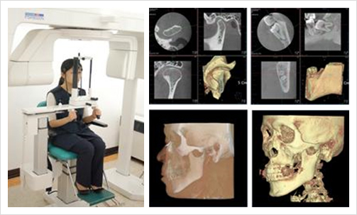
Radiation defense
The main purpose of wearing protective clothing is to protect the patient's main body parts from scattered radiation. Scattered radiation generated during intraoral radiographs or panoramic radiographs is very low and the effect on the body is negligible. However, it is a rule to use protective clothing for children and pregnant women, and this department makes it mandatory for all patients to wear protective clothing.The Department of Imaging Dentistry provides customized diagnosis for each individual, establishes a professional and systematic diagnosis system that reads various dental diseases such as CBCT and multi-image reconstruction images, and helps with post-operative management. In addition, with excellent medical staff and state-of-the-art equipment, we are focusing on optimizing radiation with excellent usefulness and effectiveness and improving safety while rapidly and accurately detecting diseases. The Department of Imaging Dentistry also thoroughly responds to radiation, and we have equipment to protect the body, such as lead-free radiation protective clothing for the head and thyroid gland, and have defense facilities that completely block radiation that can be dangerous to children or pregnant women.
Medical Fields
Bone disease imaging
Jaw joint imaging
Maxillary sinus imaging
Implant Imaging Diagnosis
Salivary gland imaging
Tumor and infection imaging diagnosis
Special imaging diagnosis (Ultrasound, Cone Beam CT)
Interventional salivary angiography and irrigation